Cataracts are part of the natural aging process. Everyone gets them to one degree or another if they live long enough. Cataracts, as they progress, create...

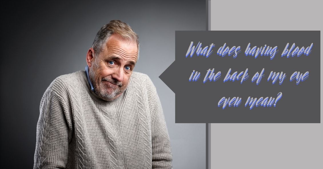
What does blood in the back of the eye signify, anyway?
It could be a retinal vein occlusion, an ocular disorder that can occur in older people where the blood vessels to the retina are blocked.
The retina is the back part of the eye where light focuses and transmits images to the brain. Blockage of the veins in the retina can cause sudden vision loss. The severity of vision loss depends on where the blockage is located.
Blockage at smaller branches in the retinal vein is referred to as branch retinal vein occlusion (BRVO). Vision loss in BRVO is usually less severe, and sometimes just parts of the vision is blurry. Blockage at the main retinal vein of the eye is referred to as central retinal vein occlusion (CRVO) and results in more serious vision loss.
Sometimes blockage of the retinal veins can lead to abnormal new blood vessels developing on the surface of the iris (the colored part of your eye) or the retina. This is a late complication of retinal vein blockage and can occur months after blockage has occurred. These new vessels are harmful and can result in high eye pressure (glaucoma), and bleeding inside the eye.
What are the symptoms of a retinal vein occlusion?
Symptoms can range from painless sudden visual loss to no visual complaints. Sudden visual loss usually occurs in CRVO. In BRVO, vision loss is usually mild or the person can be asymptomatic. If new blood vessels develop on the iris, then the eye can become red and painful. If these new vessels grow on the retina, it can result in bleeding inside the eye, causing decreased vision and floaters – spots in your vision that appear to be floating.
Causes of retinal vein occlusion
Hardening of the blood vessels as you age is what predisposes people to retinal vein occlusion. Retinal vein occlusion is more common in people over the age of 65. People with diabetes, high blood pressure, blood-clotting disorders, and glaucoma are also at higher risk for a retinal vein occlusion.
How is retinal vein occlusion diagnosed?
A dilated eye exam will reveal blood in the retina. A fluorescein angiogram is a diagnostic photographic test in which a colored dye is injected into your arm and a series of photographs are taken of the eye to determine if there is fluid leakage or abnormal blood vessel growth associated with the vein occlusion. An ultrasound or optical coherence tomography (OCT) is a photo taken of the retina to detect any fluid in the retina.
Treatment for retinal vein occlusion
Not all cases of retinal vein occlusion need to be treated. Mild cases can be observed over time. If there is blurry vision due to fluid in the retina, then your ophthalmologist may treat your eye with a laser or eye injections. If new abnormal blood vessels develop, laser treatment is performed to cause regression of these vessels and prevent bleeding inside the eye. If there is already a significant amount of blood inside the eye, then surgery may be needed to remove the blood.
Outlook after retinal vein occlusion
Prognosis depends on the severity of the vein occlusion. Usually BRVO has less vision loss compared to CRVO. The initial presenting vision is usually a good indicator of future vision. Once diagnosed with a retinal vein occlusion, it is important to keep follow-up appointments to ensure that prompt treatment can be administered to best optimize your visual potential.
Article contributed by Dr. Jane Pan
This blog provides general information and discussion about eye health and related subjects. The words and other content provided in this blog, and in any linked materials, are not intended and should not be construed as medical advice. If the reader or any other person has a medical concern, he or she should consult with an appropriately licensed physician. The content of this blog cannot be reproduced or duplicated without the express written consent of Eye IQ.